by Jerold S. Bell DVM
First printed in the July 2002 issue of the AKC Gazette
For affected dogs, hip dysplasia can be a debilitating and painful disease. It has been one of the fancy’s great challenges to combat and treat this hereditary developmental disorder, whose signs can include hip-joint pain, hind-limb weakness, lameness, exercise intolerance, degenerative joint disease, and arthritis. The disorder can include several abnormalities of the hip joints, such as joint laxity, anatomical abnormalities, and a predisposition to arthritis. While hip dysplasia is commonly perceived to be a disorder of larger dogs, it also occurs in small breeds, mixed-breed dogs, and even cats. The Pug, for example, has a significant frequency of affected dogs, while the Siberian Husky has a relatively low frequency of dysplasia.
Most experts agree that the majority of dogs that develop hip dysplasia have outwardly normal hips when they are very young, and develop the anatomical or laxity changes associated with the disorder during the first year or two of life. Initial symptoms may appear from 4 months to 1 year of age, and 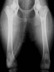 include joint pain, a swaying and unsteady gait, “bunny hopping”, difficulty rising from a sitting position, difficulty with stair-climbing, and an aggravation of these signs with exercise.
Diagnosing the Disease
There are several methods used to diagnose hip dysplasia. The standard procedure is the extended-leg (x-ray). This is a radiograph of the pelvis and hip joints taken with the dog on its back, with hind legs extended. This hip-evaluation method is the one used by the Orthopedic Foundation for Animals (OFA), as well as by most European and Canadian dysplasia-control programs.
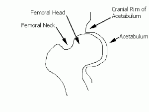
The hip is a ball (femoral head) and socket (acetabulum) joint. The OFA evaluates the hip radiograph for nine anatomical aspects. These include; a round femoral head, a deep acetabulum, a prominent notch in the femoral neck, a straight up-and-down cranial rim of the acetabulum, and minimal joint laxity. Good hip conformation is determined by imagining a line (dashed line in the drawing) connecting the outer edges of the acetabulum, and observing at least half of the femoral head enclosed within the acetabulum. Bony remodeling and arthritic changes will fill in the notch at the femoral neck, and cause “lipping” – proliferation of bone at the cranial acetabular rim.
 OFA will certify a dog’s hips (at 2 years of age or older) as excellent, good, fair, or borderline, or as mildly, moderately or severely dysplastic.
The British Veterinary Association/Kennel Club (BVA/KC) evaluates the same extended-leg radiograph at 1 year of age or older. Unlike OFA’s rating system, it separately scores the nine anatomical aspects of the pelvic radiograph for each hip. The nine scores are added up for each hip, then totaled for the dog’s final rating. A perfect rating would be zero; the worst would be 106 (53 for each hip).
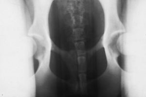 The Pennsylvania Hip Improvement Program (PennHIP) method of evaluating the hip joint is based on laxity alone. This method utilizes two separate radiographs on an anesthetized dog to record the hips in compressed and distracted views. The distracted-view radiograph (to the right) is taken while applying a uniform force on the hips to measure the maximum distractibility of the hip joints. (Distractability is the distance that the soft tissue allows the head of the femur to come out of the acetabulum.) The measured difference between the compressed and distracted views is used to compute a distraction index (DI). A DI of zero indicates no laxity of the hips, and a DI of 1.0 indicates luxation of the hips. Dogs with a DI of under 0.3 almost always have normal hips, and those over 0.7 are almost always dysplastic. PennHIP was designed to create a selection tool for tighter hips. By computing a breed average of distractibility, and selecting for tighter hips than the breed average, it is believed that the incidence of hip dysplasia should decrease over time.
Fighting Back With Breeder Selection
Hip dysplasia is considered a moderately inherited disorder, with researchers computing heritability values of 28 to 40 percent. This means that 28 to 40 percent of the variation between affected and unaffected relatives is due to genes. While it is more difficult to manage disorders with this level of heritability, many traits in this range improve with proper selection.
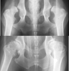 Since hip dysplasia is a polygenic disorder controlled by several gene pairs, the disease affects individual dogs due to different genetic combinations. The two dogs in the radiographs to the left have severe hip dysplasia. The one at the top has severe laxity, while the other (bottom) has tight hips, but with shallow acetabula and severe bony changes. Both these dogs have hip dysplasia, but due to different genetic causes.
One reason selection offers limited results is that many breeders are selecting dogs based solely on their OFA hip rating, and not on the specific aspects of the hip radiograph. If a dog receives a fair hip-rating due to some shallowness in it’s acetabula, it should be bred to a dog with deep acetabula (in addition to considering all other factors if it is going to be bred). If a dog’s hips have demonstrable laxity, then it should be bred to a dog with tight hips.
The BVA/KC’s dysplasia rating allows breeders to identify precisely how their dog’s hip-rating points are calculated. With this system it is easier to select prospective mates that will correct and compliment the different elements of hip joint conformation. With the OFA system, it is up to the individual breeder to work with their veterinarian to break down the hip radiograph into these separate components. By selecting for individual components of the hip radiograph, you may be more directly selecting for specific “normal-hip” genes.
The most important factor in selecting against a polygenic disorder like hip dysplasia is to seek breadth of pedigree. Most breeders select normal parents with normal 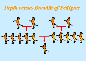 grandparents, and expect to produce all normal offspring. This is selection based on depth of pedigree. With polygenic traits, the hip status of breeding dogs’ siblings better represents the range of genes that can be present. With breadth of normalcy in the littermates of breeding dogs, and even of parents of breeding dogs, you are more efficiently selecting for a preponderance of those “normal-hip” genes.
To help control the disorder, the OFA has a longstanding hip dysplasia registry. Until recently, results were available only for dogs with normal-hip certification. The OFA has now moved to a semi-open registry, which allows owners the option of having their dogs’ hip status posted on the OFA website (www.offa.org), regardless of a normal or affected certification. The Institute for Genetic Disease Control (GDC) hip registry was developed with the open-registry concept, and, as of summer 2002, will be merged into the OFA database. Through the OFA’s online searchable registry, you can find the hip status of littermates and relatives to determine normality in the pedigree breadth. Both the OFA and PennHIP radiographic methods can report low levels of false positive and false negative predictions for future dysplastic development. The OFA radiograph documents anatomical abnormalities (shallow sockets, early bony-changes), but only natural laxity in a hip-extended view. The PennHIP radiograph documents maximum distractibility, but there are dogs with DIs between 0.3 and 0.7 that can end up being clinically normal or affected. For both methods, radiographic findings at an early age are highly correlated to dysplasia at a later age. OFA offers preliminary evaluations at any age, but does not give permanent certification until two years of age, when over 95 percent of affected dogs will show radiographic signs.
Treatment and Prevention
One significant factor in the development of hip dysplasia is the nutritional load, especially in growing large-breed dogs. Feeding high-calorie puppy food promotes rapid bone growth and weight gain. The soft-tissue components of the hips don’t mature and grow at the same rate as the bones. Often, by the time these soft-tissue components catch up, there can be bony deformity, due to a period of unstable hip joints. By switching to the lower-calorie large-breed puppy growth food, or switching to adult dog food after fourteen weeks of age, the growth rate can be slowed, and all of the components of the hip joints can mature in unison. The adult size of the dog is genetically determined, and reduced-calorie feeding will only alter the age when this size is attained.
Excessive jumping and compaction activity on the hip joints during the critical growth periods prior to skeletal maturation can also affect the degree of later dysplastic development in genetically predisposed dogs. Changing feeding protocols and managing excessive environmental stress will probably not prevent hip dysplasia in genetically predisposed dogs, just as reasonable overnutrition and activity will probably not cause hip dysplasia in genetically normal dogs. However, modifying feeding practices can alter the degree or severity of clinical signs in affected dogs. Breeders should evaluate prospective breeding dogs that have been raised under fairly uniform conditions, so that any differences between them are due to heredity, not environmental influences.
There are no scientifically proven drugs, vitamins, or food supplements that will protect the hips of genetically predisposed dogs from developing hip dysplasia. Joint-formula compounds (including glucosamine and chondroitin) are shown to diminish hip-joint pain in dogs. Nonsteroidal anti-inflammatory drugs (such as aspirin, Rimadyl, and Etogesic) are also effective for treating joint pain. Owners should discuss with their veterinarian which medications are appropriate for their dog’s condition. Avoiding overfeeding, and maintaining a lean body weight will also diminish hip pain in affected dogs.
There are two types of early-intervention surgery that attempt to prevent the progression of hip dysplasia in young dogs. These are designed to improve the integrity of the hip joints when there are shallow hip sockets or significant joint-laxity. They both act to rotate the acetabulum upward and outward, so it has greater coverage and support for the head of the femur. With a triple pelvic osteotomy (TPO), three cuts are made in the pelvis with a bone saw to isolate the hip socket. It is then rotated and reattached with metal plates. This surgery must be performed before any arthritic changes have begun.
The other surgery is an experimental procedure called a juvenile pubic symphysiodesis (JPS). An electro-scalpel is used to close the growth plate on the floor of the pelvis. With normal growth occurring in the rest of the pelvis, the hip sockets then rotate outward. This procedure must be performed on dogs between 12 and 20 weeks of age, before significant pelvic growth has occurred. The affect of the procedure beyond 2 years of age has not been studied.
Both the TPO and JPS require the early identification of candidates for surgery, before the bony changes of hip dysplasia occur. This creates a problem, as there is no accepted diagnostic test designed to predict with high certainty which dogs will develop debilitating hip dysplasia that will require surgery. Several surgeons recommend early intervention surgery based on a PennHIP measurement of laxity in young dogs. Dr. Gail Smith, professor of orthopedic surgery at the University of Pennsylvania School of Veterinary Medicine, believes that this is a misuse of the technique. “PennHIP was designed as a selection tool to quantify a probability or risk factor for developing later hip dysplasia,” says Smith, who developed the PennHIP method. “The technique wasn’t designed as an indicator for surgery.” He feels that a dog being considered for any type of hip dysplasia surgery should be demonstrating some clinical symptom of the condition.
The TPO procedure has a longer track record to measure its outcome in older dogs. While research shows that in many cases the procedure does not stop the radiographic progression of hip dysplasia or arthritis, dogs who have undergone the surgery appear to experience less discomfort. However, since studies have shown that 76 percent of all dogs with radiographic signs of hip arthritis do well without surgery, controlled studies still need to be undertaken to determine the true value of these early intervention surgeries.
There are two accepted surgical procedures for removing the pain and lameness caused by hip dysplasia: femoral head and neck excision, and total hip replacement. Both of these surgeries are considered salvage procedures, as they remove the arthritic bone-on-bone contact of the hip joint, thus relieving the pain associated with it.
A femoral head and neck excision removes the ball from the ball-and-socket joint, and uses the muscles of the pelvis to support the hind leg. This procedure works well in most dogs up to 50 pounds, though heavier dogs could also be helped. It requires good muscle strength in the leg and buttocks since these, rather than bone, provide the support.
Total hip replacement, while the more aggressive surgery, works very well on affected dogs. The head of the femur is replaced with a metal implant, and the acetabulum is replaced with a synthetic plastic implant. With updated materials and techniques, dogs receiving total hip replacement have few complications, and return to normal function without pain. Any dog whose condition is severe enough to require surgery should be spayed or neutered at the same time.
Current research
In addition to the exploration into techniques to identify and treat dogs with hip dysplasia, research is being conducted to find the genetic causes of the condition. Dr. George Brewer, of the University of Michigan Medical School, is conducting research to find genes that cause canine hip dysplasia. Using 12 breeds, he is investigating candidate genes that code for hip-related functions such as physiology and connective tissue. “We are hoping to find one or two major trigger genes for hip dysplasia in each breed,” says Brewer, whose research is supported by a grant from the AKC Canine Health Foundation.
At the Cornell University College of Veterinary Medicine, Dr. Rory Todhunter is attempting – in a different way – to identify genes that cause hip dysplasia. He bred normal Greyhounds to dysplastic Labrador Retrievers, and then bred their offspring back to either normal Greyhounds or dysplastic Labrador Retrievers. Through manipulating the genes in this breeding scheme, he is trying to identify hip dysplasia-causing genes in the normal and dysplastic crossbred offspring. The goal of both of these research efforts is to develop genetic tests that can be used to select for genetically normal breeding-dogs.
Canine hip dysplasia continues to be a serious disorder across breed lines. As breeders and owners learn the proper techniques to decrease the frequency of producing affected dogs, we can anticipate significant progress in the reduction of this damaging and costly condition.
Hip Dysplasia by Breed (OFA Statistics):
| AKC Breeds Most Affected: | AKC Breeds Least Affected: | |
| Bulldog 73.4% | Australian Terrier 0.0% | |
| Pug 60.8% | Borzoi 1.8% | |
| Otterhound 51.2% | Saluki 1.9% | |
| Clumber Spaniel 49.6% | Siberian Husky 2.1% | |
| Neopolitan Mastiff 47.6% | Ibizan Hound 2.2% | |
| St. Bernard 47.0% | Canaan Dog 2.4% | |
| Sussex Spaniel 41.2% | Pharoah Hound 2.7% | |
| Bassett Hound 28.6% | Belgian Sheepdog 2.9% | |
| Newfoundland 26.8% | Schipperke 3.0% | |
| Bloodhound 26.1% | Basenji 3.0% |
